02/14 - How Old Is Your Blood? - MSMR: What A Year!
02.14 How Old Is Your Blood?
How Old Is Your Blood?
When was the last time you tripped and fell? Perhaps you remember, perhaps you dont. As young people, toppling over in the course of your daily routine isnt something you probably worry about much. Add fifty years, however, and tripping over those shoes you left in the middle of the living room could result in serious injury.
Our bodies react differently to injury as we grow older; this is part of the normal aging process. It is an area of intense scientific inquiry because of the possibilities for preventing many age-related diseases and extending human life. Research into the aging process has often relied on experimental models in which something young is compared to something very old. Differences emerge: those differences have offered precious contributions to our growing scientific knowledge of the process of biological aging and age-associated diseases.
What is it within our bodies and genetic coding that facilitates, or perhaps permits, this transition between our time of youthful vigor and the approach of advancing age and functional decline?
Middle age
But this approach misses the time between young and old: middle age. As a middle-aged man himself, Dr. Morgan Carlson is particularly interested in this time period, when cellular processes and pathways first start to change. What is it within our bodies and genetic coding that facilitates, or perhaps permits, this transition between our time of youthful vigor and the approach of advancing age and functional decline?
Waiting until we hit our 80s or 90s to begin therapeutic intervention is not the best approach for turning back our biological clocks. But if we are able to identify and begin properly regulating age-associated determinants earlier in life, perhaps we will be able to appreciably slow down the aging process.
New research about middle age conducted by Dr. Carlson and his colleagues at the University of Connecticut Health Science Center has identified several molecular pathways that correlate with skeletal muscle aging. This research opens the possibilities for prolonging the youthfulness of our muscles well past middle age.
Skeletal muscles
are one of three major muscle types in the human body. As their name suggests, they are generally attached to bones, allowing for movement. Skeletal muscles are also known as voluntary muscles because they are under your conscious control.
To understand how skeletal muscle cells age, lets first take a look at young skeletal muscle cells.
Individual muscle cells, also known as
myocytes,
are elongated fibers with many
nuclei
that form the basis of muscle contraction. In younger bodies, myocytes are constantly regenerated from muscle stem cells that lie waiting directly outside of the mature muscle cells. These stem cells, known as
myosatellite cells,
produce new muscle cells as the mature cells are damaged or die. The transformation from myosatellite cells to myocytes is a process known as
myogenesis.
It is mediated by a variety of proteins and other signaling molecules in the body.
Dr. Carlson began his research by studying myogenesis and how it was affected by the aging process. He hypothesized that the inhibition of myogenesis observed during aging might be due to a lack of myosatellite cellsthe precursors to myocytes.
Its research you dont always get what you expect
To test this hypothesis, Dr. Carlson injected young myosatellite cells into old mice with muscle injuries. As he expected, the myosatellite cells had a remarkable effect on the injured myocytesbut not in the way he had anticipated. In fact, the injected young stem cells died within the old muscle environment. But in their death, the stem cells somehow stimulated new muscle growth such that the old injured muscle healed and actually looked younger than before.
Video of cells labeled with a probe to visualize the function of muscle stem cell mitochondria. Young or old cells were cultured in the presence of blood from mice of different ages. Level of fluorescence (red) emitted by the probe is an indicator of mitochondrial health or performance within culture environment. Results from such studies show that young muscle stem cells become metabolically dysfunctional within the presence of old blood. These findings have provided clues toward dietary regulation of age-associated molecular pathways and altered metabolic intermediates.
He found that young stem cells exposed to old blood began to act older, while old stem cells exposed to young blood acted younger there was something in the blood that was making these stem cells behave younger or older.
In order to better understand this process, Dr. Carlson pursued a series of experiments that altered the stem cell environment. In parallel experiments, Dr. Carlson exposed young stem cells to the blood of older mice and older stem cells to the blood of younger mice. He found that young stem cells exposed to old blood began to act older, while old stem cells exposed to young blood acted younger. This clearly indicated that the aging of stem cells was a matter of environment: there was something in the blood that was making these stem cells behave younger or older.
What was it?
Identifying these regenerative compounds became the mission of Dr. Carlsons research. He collected blood samples from injured and uninjured mice before and after treatment with different experimental therapies to test the levels of hundreds of proteins and other cell signaling molecules. These experiments allowed the team to home in on a protein called
transformative growth factor 1-beta,
(TGF1-β, for short).
TGF1-β plays a crucial role in regulating the
cell cycle
and in the immune response after injury. The researchers found that TGF1-β levels were significantly increased in the blood and local stem cell muscle environments of injured mice. When he compared the levels of TGF1-β in young and old mice, Dr. Carlson found that old mice had significantly elevated levels. The same was true in older humans.
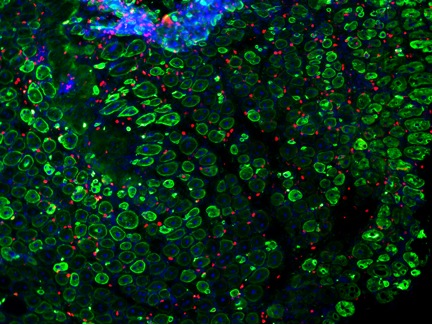
Actively regenerating skeletal muscle from a mouse. By using specific compounds like TGF1-β, it is possible to rejuvenate the regenerative abilities of muscle stem cells in older mice. Green stains newly-formed muscle fibers, red labels actively proliferating cells and blue labels all cell nuclei.
Other regenerative compounds that Dr. Carlson identified include a class of molecules called
sphingolipids.
Dr. Carlsons most recent work has focused on understanding how sphingolipids interact with the TGF1-β and other major intracellular players in the regulation of myosatellite cell aging. In addition, Dr. Carlson is studying how dietary sphingolipids and their metabolism become deregulated during aging as well as the role of the
sphinigosine-1- phosphate
pathway in the aging process.
Dr. Morgan Carlson is an Assistant Professor of Genetics and Developmental Biology at the University of Connecticut Health Sciences Center, and Principal Investigator at the UConn Stem Cell Institute. In his free time, Dr. Carlson enjoys rock climbing, pulling perfects espressos and trying to write serious pieces of contrapuntal music for the piano.
For More Information:
Loh, K. et al. 2012. Sphingosine-1-phosphate enhances satellite ell activation in dystrophic muscles through a S1PR2/STAT3 signaling pathway. PLoS One, 7(5): e37218.
Carlson, M. et al. 2009. Molecular aging and rejuvenation of human muscle stem cells. EMBO Molecular Medicine, 1: 381-391.
Carlson, M. et al. 2009. Relative roles of TGF-β1 and Wnt in the systemic regulation and aging of satellite cell responses. Aging Cell, 8: 676-689.
Carlson, M. et al. 2008. Imbalance between pSmad3 and Notch induces CDK inhibitors in old muscle stem cells. Nature, 454: 528-532.