Make a fist—this is the size of the average adult heart after it pumped out all it’s blood. Now think about the size of a tennis ball—this is the size of the average child’s heart. In newborn babies, the heart can be as small as a ping pong ball.
According to the American Heart Association, at least eight in every thousand babies is born with a heart problem at
birth,
known as a
congenital heart defect.
Some babies can grow into adults before such problems are even spotted (or become symptomatic), but some babies need immediate heart surgery to have even a chance at life. Even for heart surgeons who work with children, the small size of a newborn heart can pose many challenges for a successful surgery and long-term clinical outcomes. In some of these cases, clinicians have turned to the Visible Heart® Lab at the University of Minnesota: in this lab, founder and director Dr. Paul Iaizzo and his team can take clinical images of the patient’s heart and turn them into a 3D-printed plastic model within hours to days. These models, actual size and/or enlarged, can then be used by the clinical care team to practice the surgical procedure and also to explain the procedure and its risks to the patient’s family. “Surgeons have reported back to us that their procedures are improved as a result of studying and/or practicing with such models,” explains Dr. Iaizzo, “and also that they finally have a way to better explain to families what they are doing, why, and the risks involved.”
This is one example of the translational work happening on a daily basis within the Visible Heart® Lab, where the team of researchers, biomedical engineers, clinicians, and students seek to expand the knowledge of the heart and how this complex organ works. “Your heart beats 2 to 3 billion times in an average lifetime,” Dr. Iaizzo explains, “so why wouldn’t you want to know as much as possible about how your heart actually works?”
Dr. Iaizzo founded the Visible Heart® Lab in 1997 in collaboration with the biotechnology company Medtronic to begin studying live, large mammalian hearts in the laboratory. Over the past two decades, Dr. Iaizzo and the Visible Heart® Lab have worked to improve our understanding of human heart functional anatomy and various disease states, with a particular focus on how medical devices (such as stents, valves, catheters, pacemakers, etc.) implanted into the heart interact with native heart tissues.
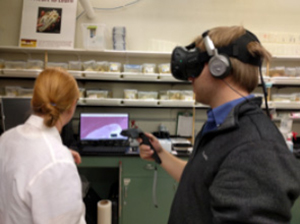
Figure 1. Mikayle Holm and Alex Mattson studying images of the heart in “Virtual Reality.”
Some of the imaging techniques used at the Visible Heart® Laboratory include: direct visualization with
endoscopes;
fluoroscopy
(x-rays);
echocardiography;
computer tomography (CT);
and
magnetic resonance imaging (MRI).
All of these imaging techniques use the physical and chemical properties in sound, light, and organic cellular matter to produce images of the relative heart anatomies and physiologic behaviors. Different techniques may be better at visualizing certain types of tissue or particular abnormalities. For example, CT images generally have better device resolution than MRI images: i.e., you can’t have most metals in an MRI. Yet, MRI images allow you to see more functional molecular details in certain tissue types. Both CT and MRI provide a fuller picture than a single x-ray. The Visible Heart® Lab atlas allows anyone to access images of the same organ created with each of these techniques to better compare them.
Video of heart 411
Reanimating Hearts
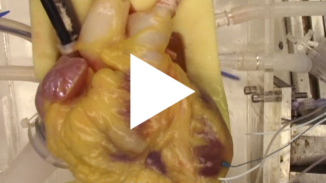
In order to better study heart functional anatomies, Dr. Iaizzo and his team have developed a method for re-animating hearts with a clear fluid (perfusate), meaning that these hearts have been removed from the body as they would be for a transplantation. But, in their lab they are working in isolation. The hearts are then attached onto a sophisticated, carefully engineered apparatus that forces fluid through the heart, which requires shocking (defibrillating) the heart into beating once again on its own.
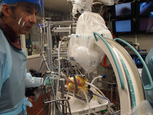
Figure 2. Dr. Iaizzo collecting multimodal images from a reanimated human heart.
Dr. Iaizzo and his research colleagues initially optimized this reanimation process on swine hearts. Then, after establishing a viable and reproducible protocol, the group began accepting human hearts that were deemed unsuitable for transplant. Over the years, Dr. Iaizzo and his team have successfully re-animated 80 human hearts, and according to Dr. Iaizzo this is one of the most amazing privileges and opportunities he has had in his research career. As Dr. Iaizzo comments: “Every human heart is unique and functionally amazing. We always learn something new anatomically or are testing new devices, maximizing what we can learn. We are always pushing the limits of the technologies we have available to us.”
Heart Video
One major goal of the lab is to improve the understanding of the general public and their appreciation of the amazing functions of the human heart: i.e., from high school students, to medical device designers, to practicing physicians. For example, the Visible Heart® Lab will print blank 3D hearts that students can then paint in color, anatomical structure by anatomical structure. As Dr. Iaizzo explains, “Once you’ve done that, you’ll never look at the human heart the same again, it puts this anatomy in your brain in a unique way.”
The same is true for the unique imaging tests that the researchers perform on the re-animated hearts, the best of which are cataloged online on the Visible Heart® Lab’s “Atlas of Human Cardiac Anatomy,” a free access website for the general public to view and utilize. “Our ultimate goal is to improve everyone’s knowledge of the human heart and how best to take care of it or treat it if such is required,” concludes Dr. Iaizzo. “These human hearts were donated to us, and we want to donate back the maximum knowledge we can gain from them.”
Dr. Paul Iaizzo is Professor of Surgery, Associate Director for the Institute for Engineering in Medicine and Founder, Director, and Principal Investigator of the Visible Heart® Laboratory at the University of Minnesota Medical School. Dr. Iaizzo first became interested in science and human physiology as a runner in college. The heart became his organ of interest because it is often the limiting factor in exercise. The Visible Heart® Laboratory is known for its collaborative nature, where students and researchers work together side by side. When not in the laboratory, Dr. Iaizzo enjoys hiking, biking, woodworking, black bear field research, and spending time with his family.
For More Information:
- Iaizzo, Paul. 2016. “The Visible Heart project and free-access website ‘Atlas of Human Cardiac Anatomy.’” Nutrition and Metabolic Insights, 5:77-83.
To Learn More:
- The Visible Heart® Lab.
http://www.vhlab.umn.edu/
- Atlas of Human Cardiac Anatomy.
http://www.vhlab.umn.edu/atlas/index.shtml
- American Heart Association.
http://www.heart.org/HEARTORG/
Written by Rebecca Kranz with Andrea Gwosdow, PhD at www.gwosdow.com