04.16 Early Detection Saves Lives - MSMR: What A Year!
04.16 Early Detection Saves Lives
Early Detection Saves Lives
Highlights
Biomaterials were used to develop a small implant for the early detection of metastatic cancer cells.
The implant captured cancer cells in mice and reduced the cancer burden in targeted organs.
The next step is clinical trials in humans to bring this device to human medicine.
Cancer treatment is all about time and space. If you find it early and treat it, you can often beat it. The early stages of cancer are localized to a specific place in the body: the breast, the colon or another organ. Over time,
cancer
grows, and spreads to other places, a process known as
metastasis.
Once cancer starts moving through the bloodstream—or metastasizing—it can be very hard to catch.
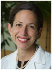
Dr. Jacqueline Jeruss is an Associate Professor of Surgery,
Pathology,
and Biomedical Engineering and Director of the Breast Care Center at the University of Michigan in Ann Arbor, Michigan. Dr. Jeruss chases cancer—particularly breast cancer—for a living. “It is so important to treat breast cancer early before it starts to spread,” Dr. Jeruss explains. But just because the tumors have been removed—with a combination of
chemotherapy,
radiation,
and surgery—the cancer can come back. Patients must continue to be tested for years after their first diagnosis, hoping to catch whatever cancer returns before it moves too far. Wouldn’t it be great if there were a better way to detect the growth of new cancer cells?
Wouldn’t it be great if there were a better way to detect the growth of new cancer cells?
This is a question that came up again and again in conversations between Dr. Jeruss and her husband Dr. Lonnie Shea, Professor and Chair of Biomedical Engineering at the University of Michigan.
Dr. Shea has used biomaterials to regenerate tissues. In this case, he adapted the material to attract cancer cells for early detection. With help from a collaborative team at the University of Michigan, Dr. Jeruss and Dr. Shea developed a small implant for the early detection of metastatic cancer.
Dr. Shea began with a
polymer
scaffolding used previously in his laboratory for delivering cells as a possible therapy for patients with
type 1 diabetes.
Since the polymer was already approved by the
Food and Drug Administration (FDA),
there is a good likelihood for translating this approach to patients.
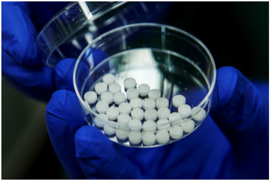
Picture of sponge-like implants
Dr. Shea implanted the small polymer device into mice with and without breast cancer and observed the types of cells captured by the implant over time. In order to identify the types of cells in the implant, Dr. Shea collaborated with the laboratory of Dr. Vadim Backman at Northwestern University in Chicago, Illinois. Dr. Backman specializes in imaging techniques and the development of non-invasive screening methods for disease diagnosis.
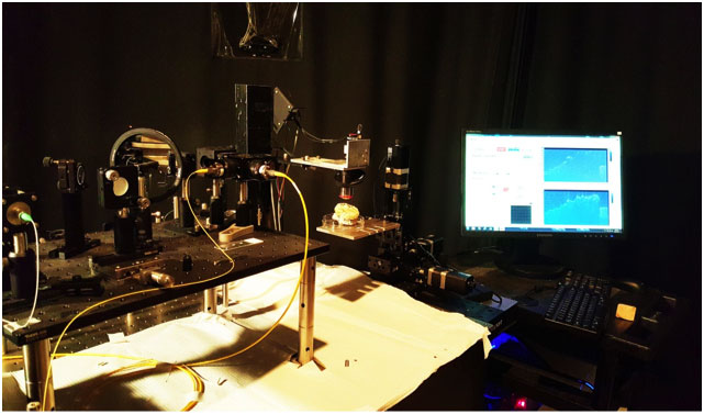
By observing the distinct ways that cancer cells alter the structure of tissue, Dr. Backman developed a method known as inverse spectroscopic optical coherence tomography (ISOCT) to detect the presence of cancer cells on the scaffold.
Picture of ISOCT machine
Using the ISOCT technique, Dr. Shea and his team found a difference in cells found in polymer implants from healthy mice and mice with breast cancer 7 and 21 days after placement. Healthy mice did not have specific immune cells in the implant. In contrast, after 7 days mice with breast cancer had many immune cells, primarily one type of immune cell called
myeloid derived suppressor cells or MDSCs.
The implant reduced the cancer burden in the targeted organs.
In addition, mice with breast cancer also had a significant amount of cancer cells in the implant itself but undetectable numbers of cells in the liver and lungs, organs that are usually targeted by metastatic breast cancer. In fact, the implant reduced the cancer burden in the targeted organs.
After 21 days, Dr. Shea and his team detected some cancer in the lungs and liver of mice with breast cancer, but the number of cells was significantly reduced compared to mice that did not receive an implant. “These results show that the polymer implant we developed provides a mechanism for early detection of metastatic cancer,” explains Dr. Shea and can reduce the cancer burden.
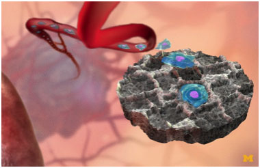
Schematic of implant capturing cancer cells
The next step is to conduct human clinical trials. Given that the implant is made from materials already approved by the FDA for use in medicine, Dr. Shea and his collaborators hope to receive permission for human clinical trials soon. The ideal candidate for the trial would be a breast cancer patient who has successfully been treated but may be at high risk for relapse. An implant in this patient could help clinicians like Dr. Jeruss better monitor their patients in a non-invasive way. “If we can show that this works in humans,” remarks Dr. Jeruss, “this would open new doors into cancer treatment, and the treatment of other diseases as well.”
Dr. Jacqueline Jeruss is Associate Professor of Surgery, Pathology, and Biomedical Engineering and Director of the Breast Care Center at the University of Michigan. Dr. Jeruss specializes in the care of patients with breast cancer or women at high risk for breast cancer. Her research interests include new methods for treating aggressive breast cancer and preventing the development of metastatic cancer.
Dr. Vadim Backman is the Walter Dill Scott Professor of Biomedical Engineering and Program Leader of the Robert H. Lurie Comprehensive Cancer Center at Northwestern University. His laboratory specializes in imaging techniques for the detection and diagnosis of disease with a focus on cancer.
Dr. Lonnie Shea is the William and Valerie Hall Chair of the Department of Biomedical Engineering and Professor of Chemical Engineering at the University of Michigan. His research focuses on the development of biomaterials for novel treatments and drug therapies.
When not in the laboratory, all three scientists enjoy spending time with their families, particularly outside.
For More Information:
Aguado BA, Caffe JR, Nanavati D, Rao SS, Bushnell GG, Azarin SM, Shea LD. Extracellular matrix mediators of metastatic cell colonization characterized using scaffold mimics of the pre-metastatic niche. Acta Biomater. 2016 Feb 1. [Epub ahead of print].
Azarin SM, Gower RM, Aguado BA, Sullivan ME, Goodman AG, Jiang EJ, Rao SS, Ren Y, Tucker SL, Backman V, Jeruss JS, Shea LD. In vivo capture and label-free detection of early metastatic cells. Nature Communications 2015:8;6:8094.