| |
 Rip Van Winkle Rip Van Winkle
According to the story by Washington Irving, a lazy man named Rip van Winkle went off into the woods to escape his wife, drank some liquor, and fell asleep for twenty years. Upon awakening, he strolled back into town to find that everything had changed. Seems plausible enough
at first
but as science students we know that after sleeping for 20 years, Rip would not have been able to walk at all. He would not even have been able to lift himself off the ground. His bone and muscle mass would have decreased to the extent that he could not support his own body weight.
People who are on bed rest or confined to a wheelchair for just a couple of months begin to lose significant bone mass. Extended periods of physical inactivity cause bone loss in other animals as well. Ground squirrels, hamsters, and little brown bats all lose bone mass during hibernation.
Loss of bone density is a problem because it leads to more easily broken bones and other complications for humans and other animals.
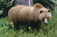
Yet somehow bears are able to maintain bone
density during winter hibernation. The key to their success may
some day help reverse bone loss in humans resulting from prolonged
periods of inactivity or from the disease osteoporosis, which
is caused by decreased bone density, and affects over half the
people over 50 years old in the U.S.
There are 256 bones in the human body that come in all different shapes and sizes. Bones are important. They provide structural support and allow for movement. They are strong enough to support your body weight; they are also the site where blood cells and bone cells are produced.
Create, Destroy, Repeat
Bones are formed from proteins such as collagen and from the mineral crystals of calcium
phosphate, which make up the bone extracellular matrix. The
bone matrix is ever-changing and the process of bone creation and destruction
goes on throughout your whole life. Two important cells responsible for these
processes are osteoblasts and osteoclasts. That one-letter difference
is huge.
Osteoblasts are responsible for bone formation.
Osteoclasts are responsible for bone destruction.
An osteoblast will create bone by secreting minerals that attach to the bone matrix proteins. Eventually these deposits need to be replenished, though, so the osteoclasts secrete enzymes that break down the mineral deposits. These minerals are then released back into the bloodstream and excreted in urine.
Bone density is determined by the rate of bone formation (osteoblast activity) compared to the rate of bone resorption (osteoclast activity). This is a continuous process called bone turnover, and your bone density will change significantly over the course of a lifetime.
In humans, from the time you are born to the ages of 20 to 30 years, bone formation exceeds bone resorption and your bone density increases. During this time, it is important to ingest minerals, particularly calcium, in order to maximize bone formation. When osteoblast activity begins to decline and bone formation no longer exceeds bone resorption, you have reached what is known as peak bone density. This is the most bone mass you will ever have. After this point, osteoclast activity begins to exceed osteoblast activity and bone density starts declining. Particularly in post-menopausal women, this decline can be so extreme that the bone structure becomes compromised. This results in weakened bones and the increased likelihood of fracture, a condition known as osteoporosis. People who do not ingest enough calcium during their years of increasing bone density put themselves at particular risk for osteoporosis.
Don't Let This Happen to You
In some people, the rate of bone resorption can prematurely exceed that of bone formation during periods of prolonged inactivity, such as a patient confined to bed rest or someone with a spinal chord injury who can no longer walk. This condition is known as disuse osteoporosis. Disuse is not only limited to these conditions. Anyone who does not get enough exercise (walking counts) puts themselves at risk for reduced bone mass and disuse osteoporosis. This can be especially harmful for children during the years of bone formation.
Do Not Try This At Home
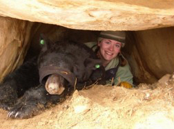 Mariah and MeghanDr. Seth Donahue of Michigan Technical University studies osteoporosis and disuse osteoporosis in bears. He decided to study bone metabolism in bears because they are physically inactive for six months of the year during hibernation. Over the course of his research, Dr. Donahue has obtained bear blood samples and bone samples from various sources. He has collaborated with researchers studying black bears in Virginia and grizzly bears in Washington State. He also obtained bear bones from hunters in Michigan and Utah. Mariah and MeghanDr. Seth Donahue of Michigan Technical University studies osteoporosis and disuse osteoporosis in bears. He decided to study bone metabolism in bears because they are physically inactive for six months of the year during hibernation. Over the course of his research, Dr. Donahue has obtained bear blood samples and bone samples from various sources. He has collaborated with researchers studying black bears in Virginia and grizzly bears in Washington State. He also obtained bear bones from hunters in Michigan and Utah.
One of Dr. Donahue's closest collaborators is Dr. Mike Vaughan, a Professor of Wildlife Biology at Virginia Technical
University. Dr. Vaughan studies the effects of human activity
on bear reproduction, physiology ,
and habitat. Through this collaboration, Dr. Donahue obtained blood
samples from both wild and captive bears and tested these samples
for hormones and markers of bone resorption and bone formation. Some of these blood samples were from pre-hibernating bears and some were from post-hibernating bears. It was thuof Dr. Donahues closest collaborators is Dr. Mike Vaughan, a Professor of Wildlife Biology ats possible to compare the hormone levels in bears pre-and post-hibernation. Based on other models of disuse osteoporosis, Dr. Donahue expected the bone resorption rate to increase. Indeed, as expected, this is what he found. However, unlike other models of disuse osteoporosis, bear bone formation rate also increased during hibernation. From these initial results, Dr. Donahue concluded that bears do not lose bone density during their periods of hibernation.
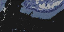 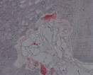 Osteoblasts in black bear bone vs. osteoclasts in black bear bone
In another experiment, he obtained large bones from bears killed during hunting season in Michigan and Utah. Hunting season in Michigan occurs once a year in the fall. Dr. Donahue compared the bones of younger and older bears to see if their bones changed with age. Bear age can be determined by growth marks on their teeth. Dr. Donahue and his team of researchers did stress tests on the bones to see how much weight each bone could hold. They also took cross-sections of the bone to measure porosity, or how many spaces were in the bones. Dr. Donahue found that the bones of older bears were actually stronger and less porous than the bones of younger bears. This is in contrast to humans, where bone strength decreases and bone porosity increases with age. Somehow the bears maintain bone formation with age despite annual periods of inactivity.
Research Is Collaboration
In Utah there are two hunting seasons: one in the
spring and one in the fall. Dr. Hal Black at Brigham
Young University provided bones from both seasons, and Dr. Donahue
was able to compare the bones of bears pre-hibernation (fall) and post-hibernation
(spring). He found that the bone structure, strength, and mineral concentration
were similar in pre- and post-hibernating bears. These results support
his previous findings that bears do not experience disuse osteoporosis
during hibernation. In order to study bone turnover more in-depth, Dr.
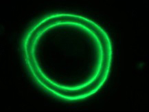 A fluorescently labeled bone remodeling unit in a grizzly bear femurDonahue collaborated with Dr. Charlie Robbins, a Professor of zoology
and Natural Resource Sciences at Washington State University. Dr. Robbins
studies the foraging ecology of bears and their role in moving nutrients
throughout the environment. The bears were given a fluorescent dye
that interacts with calcium, so when calcium is incorporated into the
bone, a fluorescent marker is attached. When these bones are put under
a fluorescent microscope, it is possible to quantify the rate of bone
formation. Some of the bones Dr. Donahue studied were from bears that
died during hibernation; some of the bones were from bears that died
after hibernation. In this way it was possible to compare rates of bone
formation in hibernating and post-hibernating bears. Dr. Donahue found
that the rate of bone formation was decreased in hibernating bears compared
to post-hibernating bears. This result was expected because metabolic
processes slow during hibernation to conserve energy. But in bears the
rates of bone resorption and bone formation remained balanced. A fluorescently labeled bone remodeling unit in a grizzly bear femurDonahue collaborated with Dr. Charlie Robbins, a Professor of zoology
and Natural Resource Sciences at Washington State University. Dr. Robbins
studies the foraging ecology of bears and their role in moving nutrients
throughout the environment. The bears were given a fluorescent dye
that interacts with calcium, so when calcium is incorporated into the
bone, a fluorescent marker is attached. When these bones are put under
a fluorescent microscope, it is possible to quantify the rate of bone
formation. Some of the bones Dr. Donahue studied were from bears that
died during hibernation; some of the bones were from bears that died
after hibernation. In this way it was possible to compare rates of bone
formation in hibernating and post-hibernating bears. Dr. Donahue found
that the rate of bone formation was decreased in hibernating bears compared
to post-hibernating bears. This result was expected because metabolic
processes slow during hibernation to conserve energy. But in bears the
rates of bone resorption and bone formation remained balanced.
What Does It Mean?
When the rate of bone resorption exceeds that of bone formation, the excess calcium and other minerals are secreted into the blood stream and excreted from the body via urination. During hibernation bears do not urinate, but the concentration of calcium in the blood must remain the same; otherwise, high blood calcium levels (hypercalcemia), can cause a variety of problems. These problems include depressed reflexes and confusion. Hypercalcemia can also affect the way cells in the body function, especially the heart. Dr. Donahue hypothesized, then, that the need to preserve bone density was driven by the need to maintain normal blood calcium levels. He believes that bears have a recycling mechanism that other animals dont have.
A human patient confined to bed rest shows an increased calcium excretion whereas a bear can maintain homeostasis during six months of hibernation. For this reason, Dr. Donahue became interested in hormones that regulate calcium. Particularly parathyroid hormone (PTH).
To test PTH concentrations in bears, Dr. Donahue used blood samples from black bears housed at Virginia Tech University. Blood samples were taken every ten days from the beginning of October to the end of May. Hibernation lasted from the beginning of January to the beginning of April. This time frame, then, encompassed pre-hibernation, hibernation, and post-hibernation periods. Tests already existed to determine PTH concentration in mice, rats, and humans. In these tests, blood samples are combined with an antibody that will react only with the specific molecule, in this case PTH. The researchers found that PTH levels increased during hibernation and were correlated with osteocalcin, a marker of bone formation that is found in the bone matrix. To Dr. Donahue, these results suggested that bear PTH might be able to stimulate osteoblast activity in people.
 A grizzly bearThen he collaborated with a pharmaceutical company to create a new drug based on bear PTH. To test the hypothesis, bear PTH was given to female rats whose ovaries had been removed. This is a model of post-menopausal osteoporosis in rats. When the ovaries are removed, the rats rapidly begin to lose bone mass. The research rats were allowed to lose bone mass for 6 weeks. They were then given weekly injections of bear PTH for eight weeks and their bone mass increased. This result was not too surprising, said Dr. Donahue, because human PTH will do the same thing. We were hoping that bear PTH would be more potent. So far, the evidence is inconclusive as to whether bear PTH is more effective at inducing bone formation than human PTH. He is hopeful that bear PTH will some day be a useful drug for patients who have lost bone mass. This includes post-menopausal women with osteoporosis, as well as older men with osteoporosis, people with spinal cord injuries, or other situations where disuse osteoporosis occurs. A grizzly bearThen he collaborated with a pharmaceutical company to create a new drug based on bear PTH. To test the hypothesis, bear PTH was given to female rats whose ovaries had been removed. This is a model of post-menopausal osteoporosis in rats. When the ovaries are removed, the rats rapidly begin to lose bone mass. The research rats were allowed to lose bone mass for 6 weeks. They were then given weekly injections of bear PTH for eight weeks and their bone mass increased. This result was not too surprising, said Dr. Donahue, because human PTH will do the same thing. We were hoping that bear PTH would be more potent. So far, the evidence is inconclusive as to whether bear PTH is more effective at inducing bone formation than human PTH. He is hopeful that bear PTH will some day be a useful drug for patients who have lost bone mass. This includes post-menopausal women with osteoporosis, as well as older men with osteoporosis, people with spinal cord injuries, or other situations where disuse osteoporosis occurs.
Wonderful Bears
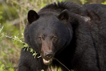 A black bearDr. Donahue also emphasizes other remarkable adaptations of hibernating bears. The dont lose bone mass during hibernation; they also dont lose much muscle mass or experience heart failure or kidney failure, as humans would under hibernating conditions. There are sophisticated mechanisms at work in bears during hibernation that act in an integrated fashion to preserve tissue. Dr. Donahue believes this is not just a case of adaptation to hibernation because other hibernating animals are not able to maintain bone and muscle mass like bears do during hibernation. A black bearDr. Donahue also emphasizes other remarkable adaptations of hibernating bears. The dont lose bone mass during hibernation; they also dont lose much muscle mass or experience heart failure or kidney failure, as humans would under hibernating conditions. There are sophisticated mechanisms at work in bears during hibernation that act in an integrated fashion to preserve tissue. Dr. Donahue believes this is not just a case of adaptation to hibernation because other hibernating animals are not able to maintain bone and muscle mass like bears do during hibernation.
Dr. Donahue has also begun to look at osteoblast activity in bears. In particular, he is studying the rate of cell death, in osteoblast cells. To do this he has incubated blood from bears with mice osteoblast cells in culture. Blood samples were taken from pre-hibernating, hibernating, and post-hibernating bears and osteoblast apoptosis activity was compared at these different times. The most interesting finding so far is that osteoblasts cultured with blood from hibernating bears have the lowest rate of apoptosis. Dr. Donahue suspects that there are circulating molecules that are protecting the osteoblasts from dying during hibernation. “The next step is to figure out what is changing between pre-hibernation and hibernation, so we can see what factors are likely to contribute to the preservation of bone cells and bone mass.”
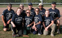 The Boneheads Softball TeamDr. Donahue is a Professor of Biomedical Engineering at Michigan Technical University. He studies hibernating bears as a model of disuse osteoporosis. He played football during college and was interested The Boneheads Softball TeamDr. Donahue is a Professor of Biomedical Engineering at Michigan Technical University. He studies hibernating bears as a model of disuse osteoporosis. He played football during college and was interested
in sports biomechanics and optimizing athletic performance. He went to graduate school in the field
of orthopedic mechanics. Dr. Donahue still loves sports and being outside. When not in the lab, he can be found on the softball field with his lab team, The Boneheads, playing hockey, downhill skiing, mountain biking, hunting, and fishing.
For more information:
- McGee-Lawrence, M. et al. Grizzly bears (Ursus arctos horribilis) and black bears (Ursus americanus) prevent trabecular bone loss during disuse (hibernation). 2009. Bone, 2009 Aug 21. [Epub ahead of print].
- McGee-Lawrence, M. et al. “Six months of disuse during hibernation does not increase intracortical porosity or decrease cortical bone geometry, strength, or mineralization in black bear femurs.” 2009. Journal of Biomechanics, 42: 1378-1383.
- McGee, M. et al. “Decreased bone turnover with balanced resorption and formation prevent cortical bone loss during disuse (hibernation) in grizzly bears.” 2008. Bone, 42: 396-404.
- Donahue, S. et al. “Parathyroid hormone may maintain bone formation in hibernating black bears to prevent disuse osteoporosis.” 2006. Journal of Experimental Biology, 209: 1630-1638.
- Donahue, S. et al. Hibernating bears as a model for preventing disuse osteoporosis. 2006. Journal of Biomechanics, 39: 1480-1488.
To learn more:
Bear Hibernation:
- Dr. Donahues website at Michigan Technical Institute Biomedical Engineering. http://www.biomed.mtu.edu/swdonahu
- Very interesting article on bear hibernation - Public Broadcasting Service http://www.pbs.org/wgbh/nova/satoyama/hibernation.html
Physical Inactivity and Bone Mass in Children:
- McKay H, Smith E. “Winning the battle against childhood physical inactivity:
the key to bone strength?” 2008. Journal of Bone Mineral Research, 23:980-5.
- Sardinha LB, et al. “Objectively measured physical activity and bone strength in 9-year-old boys and girls.” 2008. Pediatrics, 122:e728-36.
Written by Rebecca Kranz with Andrea
Gwosdow, Ph.D. Gwosdow
Associates
HOME | ABOUT | ARCHIVES | TEACHERS | LINKS | CONTACT
All content on this site is © Massachusetts
Society for Medical Research or others. Please read our copyright
statement — it is important. |
|
|
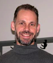
Dr. Seth Donahue
Sign Up for our Monthly Announcement!
...or  subscribe to all of our stories! subscribe to all of our stories!

What A Year! is a project of the Massachusetts
Society for Medical Research.
|
|
